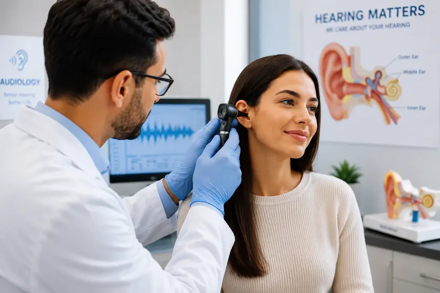
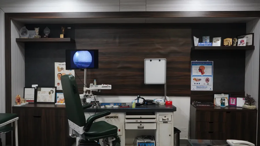
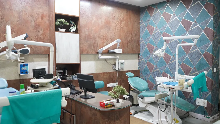
Expert ENT, Dental &
Cosmetology Under One Roof
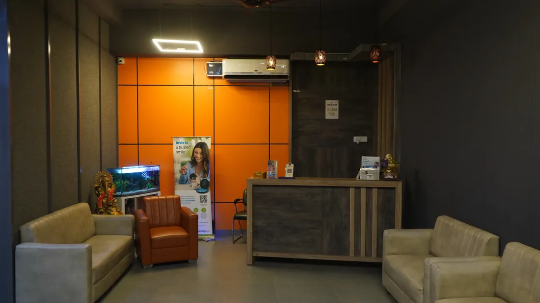
Receive expert, multidisciplinary medical and aesthetic care for the entire family in a modern, welcoming clinic located in Sector 57, Gurgaon.
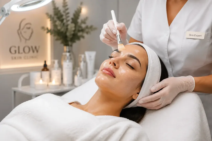
Complete Care for Every Sense
Advanced, safe treatments tailored precisely to your medical and aesthetic needs.

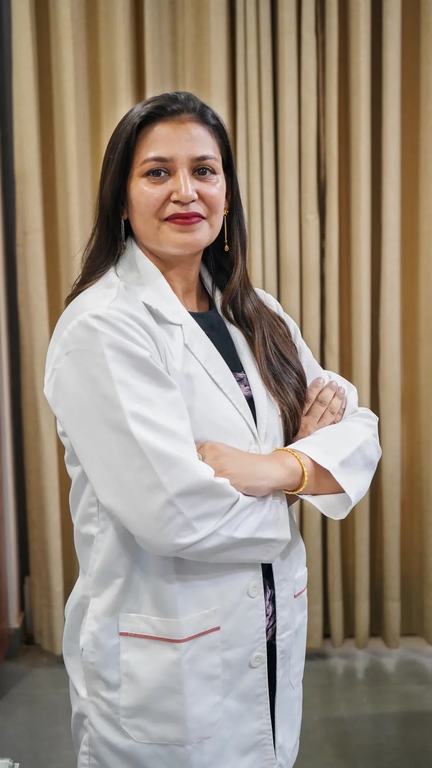
Find the Right Doctor
at Your FingertipsFind the Right Doctor Right
at Your Fingertips
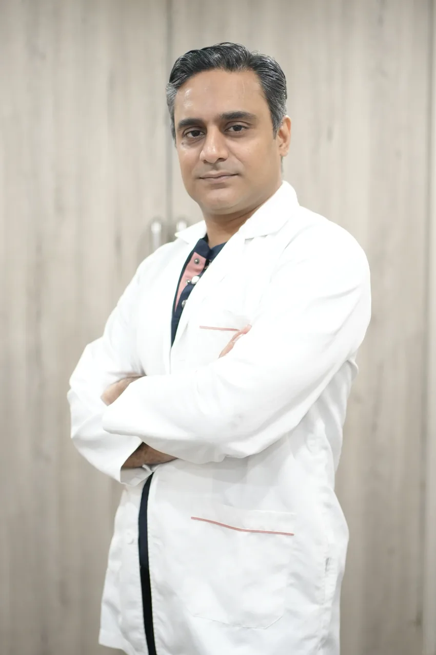
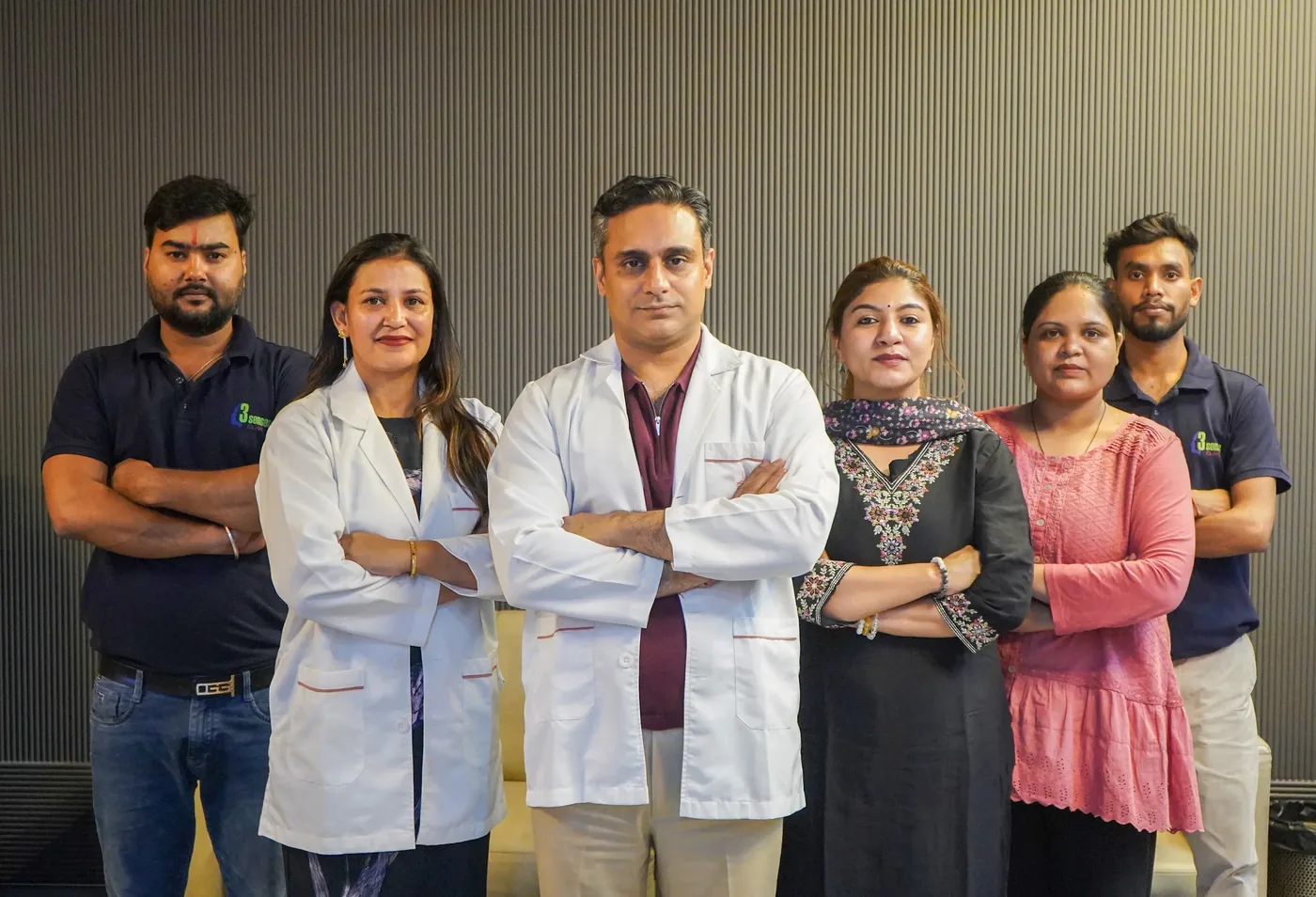
Founded by Dr Anish Gupta and Dr Priya Gupta, our facility combines medical, dental, and aesthetic expertise under one roof for seamless healthcare.
Complete Healthcare
Head, neck, and dental wellness managed in one highly convenient location.
Advanced Diagnostics
Modern medical tools utilised for precise testing and perfectly safe procedures.
Prompt Attention
Efficient scheduling ensures minimal waiting times for every single patient.
A Stellar Team of SpecialistsAlong with a Stellar Team of Specialists
Expert care led entirely by highly trained and licensed medical professionals.
Ethical Care Because Health Deserves the Best
Ethical, transparent, and gentle medical care for the Gurgaon community.
Patient Security
Strict sterilisation protocols and daily hygiene measures maintain a perfectly safe environment.
Streamlined Process
Efficient booking systems and clear consultation workflows ensure prompt medical care.
Continuous Support
A highly responsive care team remains available to answer post-treatment questions.
Verified Doctors
All treatments are provided by experienced professionals actively practising in their respective fields.
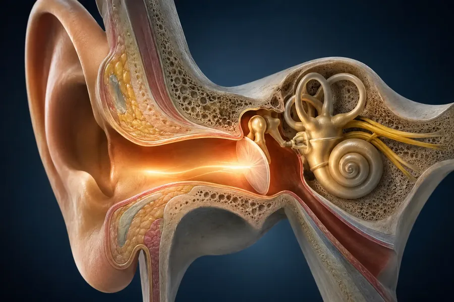
Best ENT & Dental Care
Provider Near You
Do not let health or cosmetic concerns disrupt your life. Schedule your visit to our Sector 57 clinic today.
Our Location
Sector 57, Gurgaon, Haryana, India
Email Us
info@3sensesclinics.com
Clinic Hours
Mon – Sat: 9:00 AM – 8:00 PM
Sunday: By Appointment Only
Book Your Appointment
Fill the form — we'll confirm your slot within minutes.
Also Check Out Our Videos
Educational clinical insights directly from leading specialists.
Latest Health Insights
Medical articles and expert tips to support your daily wellness.